Digital radiography is the modern method dentists use to produce X-ray images using electronic sensors and computer processing. Unlike traditional film, digital systems capture images as digital files that can be viewed, enhanced, and stored almost instantly. For patients, that translates into streamlined appointments, quicker answers, and a clearer explanation of what the dentist sees and why a particular treatment is recommended.
Because images appear on-screen immediately, clinicians can review them with patients in real time, pointing out areas of concern and comparing views side-by-side. This visual collaboration helps patients understand diagnoses and treatment options without requiring lengthy waits for film development. The immediacy also supports more efficient record keeping and coordination across the care team.
At Riverbend Family Dental we prioritize technologies that improve diagnostic clarity while keeping patient experience at the forefront. Digital radiography is one of those tools — it integrates seamlessly into modern workflows and supports better communication between clinicians and the people they treat.
Digital sensors are designed to detect X-ray photons and convert them into electrical signals that a computer can translate into a detailed image. Unlike film, which relies on a chemical reaction, digital detectors respond quickly and consistently to exposure, producing images with high contrast and sharpness. Clinicians can apply software adjustments to highlight subtle differences in density, which often makes it easier to spot early-stage problems such as small cavities or beginning bone loss.
Because digital images can be magnified and manipulated without degradation, dentists can examine suspicious areas more closely and from different angles. This ability to zoom in and adjust contrast or brightness reduces the likelihood of missing fine details and supports more precise treatment planning. The diagnostic advantages are particularly valuable for areas that are difficult to see with a visual exam alone.
Digital radiography also supports advanced imaging workflows, such as combining intraoral sensor images with panoramic or 3D scans when a more comprehensive view is needed. The compatibility between these systems promotes a cohesive diagnostic picture without requiring repeat exposures or awkward transfers of physical film.
One of the most practical benefits of digital radiography is speed: images are available within seconds, enabling faster clinical decisions. This reduces chair time and can shorten the interval between diagnosis and treatment. When a problem is identified, the dental team can immediately begin to outline next steps, discuss conservative options, and explain anticipated outcomes using the same images the clinician is viewing.
Speed also improves coordination when multiple practitioners need to consult on a case. Digital files can be shared securely with specialists or sent for second opinions without the logistical delays associated with physical film. That ease of sharing supports timely referrals and helps ensure treatment plans are informed by appropriate expertise when required.
From the patient’s perspective, faster diagnosis means more predictable visits and less uncertainty. Clinicians can respond to clinical findings in the moment, recommending appropriate interventions or monitoring strategies without relying on delayed test results.
Digital radiography often improves patient comfort because the sensors are generally thinner and more flexible than older film holders. Newer sensor designs reduce gag reflexes and allow more comfortable positioning, which is especially helpful for people who experience anxiety or sensitivity during intraoral procedures. Shorter exposure times and fewer repeat images also make the process less burdensome for patients.
Beyond comfort, the ability to show patients their own images contributes to informed decision-making. When clinicians can annotate and display images on a monitor, patients gain a clearer understanding of their oral health status. Visual aids help demystify clinical terms and make it easier to explain why certain treatments are recommended or why monitoring is the best course in a particular situation.
Good communication built around clear images fosters trust and encourages patients to participate actively in their care. It also supports preventive efforts by making early issues visible before they progress into more complex problems.
Digital radiography eliminates the need for chemical developers and film processing, which has environmental benefits and reduces the amount of hazardous waste produced by a dental practice. By removing film and paper from the workflow, dental offices can operate with a smaller ecological footprint while still maintaining high diagnostic standards.
Operationally, digital systems simplify record keeping and storage. Digital images are easier to index, backup, and retrieve than physical film, reducing the risk of lost or damaged records. This streamlined approach improves practice efficiency and helps ensure continuity of care when images need to be reviewed months or years after they were taken.
In addition, digital files can be integrated into electronic health records and imaging software, enabling more robust analytics and longitudinal tracking of a patient’s oral health. These capabilities are valuable for monitoring changes over time and for creating targeted prevention strategies based on an individual’s imaging history.
While technology evolves, the underlying advantage remains: digital radiography supports high-quality diagnostics, smoother administrative processes, and a more sustainable approach to imaging compared with traditional film-based methods.
Summary: Digital radiography brings faster, clearer, and more environmentally responsible imaging to routine dental care. By combining sensitive sensors with immediate image processing, this technology enhances diagnosis, simplifies communication, and helps clinicians deliver care with greater precision and comfort. If you have questions about how digital radiography is used in our practice or how it might affect your next appointment, please contact us for more information.


Digital radiography uses electronic sensors and computer processing to produce dental X-ray images as digital files rather than film. Unlike traditional film, digital systems deliver images almost instantly, enabling clinicians to view, enhance and store images immediately. For patients, that means shorter waits, clearer visual explanations and more efficient appointments.
Digital files can be magnified, adjusted for contrast and compared side-by-side, which helps both clinicians and patients understand clinical findings. They support fast record keeping and seamless integration with electronic health records for long-term tracking of oral health. Overall, digital radiography streamlines diagnostic workflows and improves communication during treatment planning.
Digital radiography exposes patients to a fraction of the radiation used by older film techniques because digital detectors are more sensitive and require shorter exposures. Modern dental X-ray equipment and positioning techniques further reduce exposure while maintaining image quality. Practices follow radiation safety principles such as ALARA and use shielding and careful technique to limit dose.
If you are pregnant or believe you might be, inform the dental team so they can take additional precautions or postpone nonurgent imaging. Lead aprons, thyroid collars and judicious imaging choices are common safeguards to protect sensitive tissue when X-rays are necessary. Your clinician will balance diagnostic benefit and safety when recommending any radiographic study.
Digital sensors detect X-rays and convert them into electrical signals that a computer translates into detailed images with high contrast and sharpness. Because detectors respond consistently to exposure, clinicians can rely on predictable image quality and reduce repeat exposures. Software tools allow adjustment of brightness, contrast and magnification to reveal subtle changes in tooth and bone structure.
The ability to enlarge and manipulate images without degradation helps clinicians identify early decay, bone changes and other fine details that are difficult to see visually. Digital images also integrate with other imaging modalities, allowing clinicians to combine intraoral images with panoramic or 3D scans when a broader view is needed. This interoperability supports more complete diagnoses without unnecessary repeat imaging.
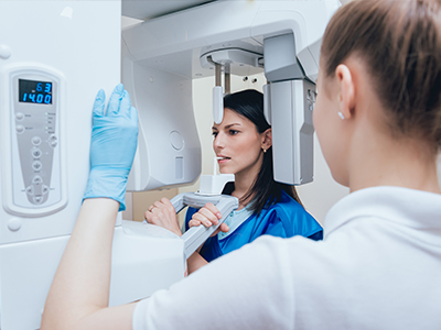
During an appointment, the dental assistant or hygienist will position a thin intraoral sensor and take a few quick exposures while you remain seated. Each exposure lasts only seconds, and images appear on the monitor almost immediately for review by the clinician. This immediate feedback lets your dental team explain findings, answer questions and discuss next steps while you are still in the chair.
Sensors are smaller and more flexible than older film holders, which often improves comfort and reduces the need for repeat images. If you have a sensitive gag reflex or dental anxiety, the team can use child-sized sensors, different positioning techniques or extra time to help you feel more comfortable. Clear images shown on screen also help you make informed decisions about preventive care and treatment options.
Instant access to high-quality images speeds clinical decision-making and supports more precise treatment planning for restorations, root canal therapy and periodontal care. Digital enhancements can make small cavities, hairline fractures and early bone loss easier to detect than with unaided visual exams. This earlier detection often allows clinicians to recommend conservative measures before problems progress.
At Riverbend Family Dental, clinicians use digital radiography alongside clinical exams to build a complete diagnostic picture and tailor care to each patient. The ability to compare current images with past records helps monitor changes over time and refine treatment strategies. Digital images also facilitate timely referrals when specialist input is needed, improving coordination of care.
Digital images are stored as encrypted files within the practice’s electronic records system and included in routine backups to protect against loss. Secure archiving makes it easy for clinicians to retrieve prior images for comparison and continuity of care. Access is limited to authorized staff in accordance with privacy regulations to maintain patient confidentiality.
When specialist consultation is necessary, images can be transmitted through secure, HIPAA-compliant channels to minimize delays. Patients may also request copies of their images for second opinions or for transfer to another provider, and the practice will follow secure procedures for release. These secure workflows reduce administrative friction while preserving privacy and clinical utility.
Because modern sensors are thinner and come in a range of sizes, many patients find intraoral imaging more comfortable than older film systems. Shorter exposure times and fewer repeat images further reduce discomfort and the overall time required for imaging. Clinicians can modify positioning and use bite blocks or foam supports to help patients who have difficulty holding sensors in place.
Pediatric patients benefit from smaller sensors and techniques designed for children, which improve comfort and cooperation during imaging. For adults with strong gag reflexes, staff may use distraction, adjusted angulation or additional support to complete studies with minimal distress. Comfort-focused imaging practices help ensure diagnostic quality while supporting a positive patient experience.
Digital radiography eliminates film, chemical developers and associated hazardous waste, reducing the environmental footprint of dental imaging. Removing chemistry from the workflow also simplifies infection control and waste handling within the office. Many practices find digital systems reduce paper use and physical storage needs as images are kept electronically.
Digital workflows support easier record management, indexing and long-term tracking without the need for physical archives. These operational efficiencies contribute to sustainability goals while maintaining high diagnostic standards. Overall, the move to digital imaging aligns environmental responsibility with improved clinical capabilities.
Intraoral digital sensors excel at detailed images of individual teeth and surrounding bone but do not replace panoramic or 3D cone-beam scans when broader anatomic context is required. Panoramic imaging provides a wide view of jaws and dentition, and CBCT delivers three-dimensional detail for implant planning, complex extractions and airway assessment. Clinicians select the modality that best answers the diagnostic question while minimizing exposure.
Many modern practices integrate intraoral, panoramic and 3D imaging into a cohesive workflow so that each tool contributes the most useful information. At Riverbend Family Dental, this integrated approach helps clinicians choose the least invasive option that still provides the necessary diagnostic detail. When advanced imaging is indicated, clinicians will explain why it is recommended and how it complements intraoral digital radiography.
There is no universal schedule for dental X-rays; frequency depends on individual risk factors such as age, oral health history and current clinical findings. Patients with a history of decay, active periodontal disease or ongoing dental treatment may require images more often than those with healthy, stable mouths. Clinicians assess risk at each visit and recommend imaging only when it will influence diagnosis or management.
Digital radiography makes comparisons across visits quick and informative, which can support longer intervals between images when appropriate. If you have questions about why a particular image is recommended, your dental team will explain the clinical rationale and how the result will guide care. Open communication ensures imaging decisions align with your health needs and personal preferences.
Ready to transform your dental experience?
At Riverbend Family Dental, our team makes achieving optimal oral health easy and stress-free. From routine check-ups to treatments like Invisalign, we provide clear communication and patient-focused care every step of the way.
Getting started is simple—call, email, or use our online portal to schedule a visit and have your questions answered by our experts. Don’t wait—contact Riverbend Family Dental today and experience compassionate, precise care for your smile.