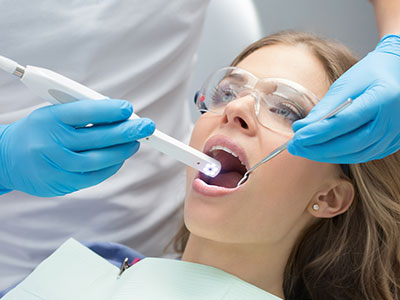
Intraoral cameras bring the inside of the mouth into clear view for both clinicians and patients. These compact, pen-sized devices capture high-resolution, full-color images of teeth, gums and other oral structures and display them instantly on a monitor. At Riverbend Family Dental, we use intraoral imaging to make examinations more transparent, improve diagnostic accuracy, and involve patients directly in decisions about their oral health.
Intraoral cameras are designed to illuminate and magnify areas that are difficult to inspect with the naked eye. Cracks in enamel, hairline fractures, early decay along margins, worn restorations and subtle changes in soft tissue all become easier to identify when viewed at high magnification. The color fidelity and sharpness of modern devices allow clinicians to detect variations that suggest early-stage problems rather than relying solely on tactile feedback or visual inspection.
Beyond hard-tissue issues, these cameras can document soft-tissue conditions such as inflamed gingiva, suspicious lesions, or areas of recession. Because the images are recorded in real time, clinicians can rotate the camera, change angles and highlight specific regions immediately — a capability that improves the thoroughness of the exam and helps locate issues that might otherwise be missed.
Captured images can be reviewed frame-by-frame, which is particularly useful when monitoring progressive changes over time. This visual archive supports more informed decisions about when to intervene and when to monitor conservatively, offering a clearer clinical picture than notes alone.
One of the most important benefits of intraoral cameras is their role as a communication tool. Patients often struggle to appreciate the details of an oral condition described verbally; seeing a magnified, color-accurate photograph on screen makes the condition immediately comprehensible. This shared visual context helps patients ask informed questions and feel more confident about recommended care options.
Presenting images during the consultation also reduces uncertainty and strengthens the clinician-patient partnership. When patients see the same photos the dentist is using to explain findings, there is less room for misinterpretation. This transparency supports clearer consent conversations and fosters a collaborative approach to care planning.
Because images can be annotated or compared side-by-side, clinicians can demonstrate how a condition has progressed or responded to previous treatment. That educational aspect empowers patients to take meaningful steps in preventive care and to recognize early signs that warrant attention between visits.
Intraoral cameras play a central role in sharpening diagnoses. High-resolution imaging helps confirm clinical suspicions and provides objective evidence that complements other diagnostic tools, such as radiographs. For example, an intraoral photograph can illustrate a marginal breakdown around an existing filling or reveal a fracture line that warrants a different restorative approach.
When it comes to treatment planning, these images become part of a comprehensive record that informs restorative choices, material selection and sequencing of treatment. Photographs can be shared with dental laboratories to ensure accurate shade matching and restoration fit, or with specialists to communicate precise findings before referrals. This reduces ambiguity and speeds collaborative workflows.
From a record-keeping standpoint, intraoral images are permanent additions to the patient chart. They create an audit trail of oral health over time and can be included in clinical reports or insurance documentation when needed. Because they are objective and reproducible, these images increase the clarity of clinical notes and help protect both patient and provider interests.
Intraoral cameras are lightweight, easy to disinfect and compatible with most contemporary practice management and imaging systems. Integration typically involves pairing the camera with chairside software so images appear on the monitor as they are captured and are stored automatically in the patient record. This near-instant capture-to-chart flow minimizes administrative burden and keeps clinical encounters efficient.
Clinicians use intraoral imaging at multiple points in a visit — during routine exams, pre-procedural assessments and post-treatment checks. Because the devices are non-invasive and quick to use, they fit seamlessly into standard clinical protocols without adding significant time to appointments. Many practices find that the added clarity actually streamlines consultations by resolving questions immediately.
For staff, standardized imaging protocols improve consistency. When team members follow the same sequence of views and labeling conventions, it becomes easier to retrieve and compare images across visits. This operational consistency supports better long-term monitoring and helps clinicians maintain high standards of care.
Intraoral cameras are designed with patient comfort in mind. They are small, lightweight and maneuverable, so capturing a full set of diagnostic images is typically quick and well tolerated. The devices rely on visible light and high-resolution sensors — there is no radiation exposure from intraoral photography itself, making it a safe adjunct to other diagnostic methods.
Before imaging begins, clinicians explain the process and position the camera to minimize any gag reflex or discomfort. The procedure is passive for the patient: the clinician manipulates the camera while the patient simply opens and closes their mouth as instructed. For anxious patients, the visual element can be reassuring, as they can see the examination progress on screen and understand what the provider is observing.
After images are taken, clinicians review notable findings with the patient, annotating or enlarging specific views as needed to clarify concerns. These documented images can then inform follow-up plans, monitoring schedules or referrals, and they remain available for review in future visits.
Intraoral cameras are a straightforward yet powerful technology that enhances diagnosis, improves patient communication and strengthens clinical documentation. If you’d like to learn more about how this tool is used in routine exams and treatment planning, please contact us for more information.


An intraoral camera is a compact, pen-sized digital camera that captures high-resolution images of the teeth, gums and other soft tissues inside the mouth. The device uses visible light and a high-quality sensor to produce color-accurate photographs that are displayed on a chairside monitor in real time. Clinicians can rotate and angle the camera to record multiple views, enabling close inspection of areas that are difficult to see with the naked eye.
Images are typically captured directly into the practice management or imaging software and attached to the patient record for review. Because the photos are instantaneous and clear, the clinician can freeze, enlarge or annotate specific frames during the exam. This immediate feedback loop helps both the clinician and patient evaluate findings more precisely than with unaided visual inspection alone.
Intraoral cameras reveal fine details such as cracks in enamel, hairline fractures, marginal decay around restorations and early soft-tissue changes that might be overlooked on routine inspection. They are particularly helpful for identifying subtle surface changes, worn restorations and discoloration that warrant closer monitoring or intervention. The color fidelity and magnification also assist in documenting inflamed gingiva, recession and suspicious lesions.
Because images can be captured from multiple angles and saved over time, intraoral photography is useful for tracking progressive conditions and evaluating treatment outcomes. Repeated images create a visual timeline that aids clinical decision-making about when to treat versus when to observe. This documentation also supports communication with specialists when a referral is needed.
High-resolution intraoral images provide objective visual evidence that complements tactile examination and radiographs, helping clinicians confirm clinical suspicions with greater confidence. Photographs can highlight marginal breakdown around fillings, fracture lines or early decay that may alter the recommended restorative approach. The clarity of the images improves material selection, shade matching and the sequencing of procedures during treatment planning.
Images are easily included in the patient chart and shared with laboratories or specialists to ensure accurate communication about findings and expectations. This reduces ambiguity in laboratory prescriptions and specialist consults, which can improve turnaround and fit. The visual record also assists clinicians in prioritizing treatment and explaining why certain steps are recommended.
Intraoral imaging is noninvasive and generally well tolerated by patients; the cameras are small, lightweight and designed for comfortable intraoral use. The procedure involves the clinician positioning the device while the patient simply opens and closes the mouth as directed, so no active work or pressure is applied to teeth or soft tissues. Because the cameras rely on visible light only, there is no radiation exposure associated with the photographs themselves.
For patients who gag or feel anxious, clinicians use positioning techniques and gentle pacing to minimize discomfort and allow for clear images to be taken. Images are captured quickly, and clinicians can pause to explain what is being seen on screen to reassure the patient. If needed, the team will adapt their technique to make the process easier for children or those with special needs.
Intraoral photographs are typically saved directly into the digital patient record and organized alongside radiographs and clinical notes for easy retrieval. These images become part of the ongoing clinical archive and allow clinicians to review changes over time by comparing visits. Proper labeling and consistent naming conventions make it simpler to find specific views when planning care or documenting outcomes.
Stored images can be included in clinical reports, treatment plans and referral documentation when appropriate, providing objective visual evidence to support clinical decisions. The permanent record helps the care team monitor progression, verify the condition of restorations and communicate findings clearly to patients. Secure imaging systems also protect patient privacy while allowing authorized staff to access necessary files during treatment.
Intraoral cameras translate clinical observations into clear, color-accurate images that patients can see and understand, which makes explanations more concrete and less abstract. When patients view the same photographs the clinician is using, they can follow the discussion more easily and ask targeted questions about specific areas of concern. This shared visual context promotes informed decision-making and helps patients feel more engaged in their care.
At Riverbend Family Dental, clinicians use captured images to annotate findings and show progression or response to treatment during follow-up visits. Seeing side-by-side comparisons empowers patients to recognize early warning signs and take preventive steps between appointments. Clear visuals also help the dental team explain recommended procedures and expected outcomes in a collaborative way.
Yes, intraoral cameras are safe and adaptable for use with children and patients who have special care requirements because they are noninvasive and employ only visible light imaging. The devices are small and maneuverable, allowing clinicians to capture diagnostic views without causing discomfort, and clinicians often modify their approach to accommodate limited cooperation. Short imaging sessions and patient-friendly positioning make it possible to obtain useful diagnostic images even in younger or anxious patients.
Clinicians can use behavioral techniques, distraction and gradual exposure to help children tolerate the procedure, and caregivers are often invited to be present to increase comfort. For patients with mobility or sensory challenges, the team will tailor the session length and positioning to prioritize safety and dignity. Images taken during these visits still provide valuable documentation for monitoring growth, development and oral health status.
Intraoral cameras are designed to work seamlessly with chairside software, digital radiography and practice management systems so that images appear on the monitor as they are captured and are stored automatically in the chart. This integration creates an efficient capture-to-chart workflow that minimizes administrative steps and keeps clinical encounters focused on patient care. Coordinated systems also allow clinicians to view radiographs and intraoral photos side-by-side for a more comprehensive assessment.
The practice leverages these integrations to improve clinical consistency and interdisciplinary collaboration by sharing images with labs and specialists when needed. Standardized imaging protocols and labeling conventions make it easier to retrieve comparable views across visits and providers. Ultimately, the connectivity between devices reduces errors and supports a smoother patient experience from diagnosis through treatment.
Yes, intraoral photographs are a convenient way to share precise visual information with specialists and dental laboratories to support referrals and restorative work. High-quality images help laboratories match shades, assess occlusal relationships and fabricate restorations that fit more accurately, while specialists benefit from detailed documentation when planning shared care. Files are typically exported in secure formats and accompanied by clinical notes describing the relevant findings.
Sharing visual evidence ahead of a referral often speeds collaboration and reduces uncertainty about the patient’s condition by providing a clear starting point for consultation. Images can be annotated to highlight specific concerns and included with digital records to ensure consistent communication. Secure transfer methods and strict privacy protocols protect patient information during these exchanges.
Dentists will explain the purpose of the intraoral imaging and show the patient how the camera will be used before beginning the exam. The clinician will then capture a series of targeted photographs while the patient follows simple instructions to open, close or reposition the tongue as needed, which generally takes only a few minutes. After capturing images, the clinician will review notable findings on the monitor, enlarge or annotate views and discuss recommended next steps.
Patients can expect the imaging process to be quick, noninvasive and painless, with immediate visual feedback that helps clarify oral health status. The recorded images will be stored in the patient’s chart for future comparisons and care planning. If additional follow-up or specialist input is advised, the images will be used to explain the rationale and to coordinate any necessary next steps.
Ready to transform your dental experience?
At Riverbend Family Dental, our team makes achieving optimal oral health easy and stress-free. From routine check-ups to treatments like Invisalign, we provide clear communication and patient-focused care every step of the way.
Getting started is simple—call, email, or use our online portal to schedule a visit and have your questions answered by our experts. Don’t wait—contact Riverbend Family Dental today and experience compassionate, precise care for your smile.